Tag Archives: stem-cell

Good Mutations and Breathing

Van_andel_113

Stem cells — the factories that manufacture all our component body parts — may hold a key to divining why our bodies gradually break down as we age. A new body of research shows how the body’s population of blood stem cells mutates, and gradually dies, over a typical lifespan. Sometimes these mutations turn cancerous, sometimes not. Luckily for us, the research is centered on the blood samples of Hendrikje van Andel-Schipper — she died in 2005 at the age of 115, and donated her body to science. Her body showed a remarkable resilience — no hardening of the arteries and no deterioration of her brain tissue.  When quizzed about the secret of her longevity, she once retorted, “breathing”.

From the New Scientist:

Death is the one certainty in life – a pioneering analysis of blood from one of the world’s oldest and healthiest women has given clues to why it happens.

Born in 1890, Hendrikje van Andel-Schipper was at one point the oldest woman in the world. She was also remarkable for her health, with crystal-clear cognition until she was close to death, and a blood circulatory system free of disease. When she died in 2005, she bequeathed her body to science, with the full support of her living relatives that any outcomes of scientific analysis – as well as her name – be made public.

Researchers have now examined her blood and other tissues to see how they were affected by age.

What they found suggests, as we could perhaps expect, that our lifespan might ultimately be limited by the capacity for stem cells to keep replenishing tissues day in day out. Once the stem cells reach a state of exhaustion that imposes a limit on their own lifespan, they themselves gradually die out and steadily diminish the body’s capacity to keep regenerating vital tissues and cells, such as blood.

Two little cells

In van Andel-Schipper’s case, it seemed that in the twilight of her life, about two-thirds of the white blood cells remaining in her body at death originated from just two stem cells, implying that most or all of the blood stem cells she started life with had already burned out and died.

“Is there a limit to the number of stem cell divisions, and does that imply that there’s a limit to human life?” asks Henne Holstege of the VU University Medical Center in Amsterdam, the Netherlands, who headed the research team. “Or can you get round that by replenishment with cells saved from earlier in your life?” she says.

The other evidence for the stem cell fatigue came from observations that van Andel-Schipper’s white blood cells had drastically worn-down telomeres – the protective tips on chromosomes that burn down like wicks each time a cell divides. On average, the telomeres on the white blood cells were 17 times shorter than those on brain cells, which hardly replicate at all throughout life.

The team could establish the number of white blood cell-generating stem cells by studying the pattern of mutations found within the blood cells. The pattern was so similar in all cells that the researchers could conclude that they all came from one of two closely related “mother” stem cells.

Point of exhaustion

“It’s estimated that we’re born with around 20,000 blood stem cells, and at any one time, around 1000 are simultaneously active to replenish blood,” says Holstege. During life, the number of active stem cells shrinks, she says, and their telomeres shorten to the point at which they die – a point called stem-cell exhaustion.

Holstege says the other remarkable finding was that the mutations within the blood cells were harmless – all resulted from mistaken replication of DNA during van Andel-Schipper’s life as the “mother” blood stem cells multiplied to provide clones from which blood was repeatedly replenished.

She says this is the first time patterns of lifetime “somatic” mutations have been studied in such an old and such a healthy person. The absence of mutations posing dangers of disease and cancer suggest that van Andel-Schipper had a superior system for repairing or aborting cells with dangerous mutations.

Read the entire article here.

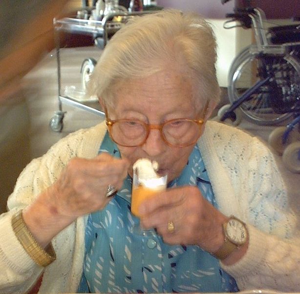
Image: Hendrikje van Andel-Schipper, aged 113. Courtesy of Wikipedia.

Growing a Brain Outside of the Body

‘Tis the stuff of science fiction. And, it’s also quite real and happening in a lab near you.

From Technology Review:

Scientists at the Institute of Molecular Biotechnology in Vienna, Austria, have grown three-dimensional human brain tissues from stem cells. The tissues form discrete structures that are seen in the developing brain.

The Vienna researchers found that immature brain cells derived from stem cells self-organize into brain-like tissues in the right culture conditions. The “cerebral organoids,” as the researchers call them, grew to about four millimeters in size and could survive as long as 10 months. For decades, scientists have been able to take cells from animals including humans and grow them in a petri dish, but for the most part this has been done in two dimensions, with the cells grown in a thin layer in petri dishes. But in recent years, researchers have advanced tissue culture techniques so that three-dimensional brain tissue can grow in the lab. The new report from the Austrian team demonstrates that allowing immature brain cells to self-organize yields some of the largest and most complex lab-grown brain tissue, with distinct subregions and signs of functional neurons.

The work, published in Nature on Wednesday, is the latest advance in a field focused on creating more lifelike tissue cultures of neurons and related cells for studying brain function, disease, and repair. With a cultured cell model system that mimics the brain’s natural architecture, researchers would be able to look at how certain diseases occur and screen potential medications for toxicity and efficacy in a more natural setting, says Anja Kunze, a neuroengineer at the University of California, Los Angeles, who has developed three-dimensional brain tissue cultures to study Alzheimer’s disease.

The Austrian researchers coaxed cultured neurons to take on a three-dimensional organization using cell-friendly scaffolding materials in the cultures. The team also let the neuron progenitors control their own fate. “Stem cells have an amazing ability to self-organize,” said study first author Madeline Lancaster at a press briefing on Tuesday. Others groups have also recently seen success in allowing progenitor cells to self-organize, leading to reports of primitive eye structures, liver buds, and more (see “Growing Eyeballs” and “A Rudimentary Liver Is Grown from Stem Cells”).

The brain tissue formed discrete regions found in the early developing human brain, including regions that resemble parts of the cortex, the retina, and structures that produce cerebrospinal fluid. At the press briefing, senior author Juergen Knoblich said that while there have been numerous attempts to model human brain tissue in a culture using human cells, the complex human organ has proved difficult to replicate. Knoblich says the proto-brain resembles the developmental stage of a nine-week-old fetus’s brain.

While Knoblich’s group is focused on developmental questions, other groups are developing three-dimensional brain tissue cultures with the hopes of treating degenerative diseases or brain injury. A group at Georgia Institute of Technology has developed a three-dimensional neural culture to study brain injury, with the goal of identifying biomarkers that could be used to diagnose brain injury and potential drug targets for medications that can repair injured neurons. “It’s important to mimic the cellular architecture of the brain as much as possible because the mechanical response of that tissue is very dependent on its 3-D structure,” says biomedical engineer Michelle LaPlaca of Georgia Tech. Physical insults on cells in a three-dimensional culture will put stress on connections between cells and supporting material known as the extracellular matrix, she says.

Read the entire article here.

Image: Cerebral organoid derived from stem cells containing different brain regions. Courtesy of Japan Times.

Building a Liver

In yet another breakthrough for medical science, researchers have succeeded in growing a prototypical human liver in the lab.

From the New York Times:

Researchers in Japan have used human stem cells to create tiny human livers like those that arise early in fetal life. When the scientists transplanted the rudimentary livers into mice, the little organs grew, made human liver proteins, and metabolized drugs as human livers do.

They and others caution that these are early days and this is still very much basic research. The liver buds, as they are called, did not turn into complete livers, and the method would have to be scaled up enormously to make enough replacement liver buds to treat a patient. Even then, the investigators say, they expect to replace only 30 percent of a patient’s liver. What they are making is more like a patch than a full liver.

But the promise, in a field that has seen a great deal of dashed hopes, is immense, medical experts said.

“This is a major breakthrough of monumental significance,” said Dr. Hillel Tobias, director of transplantation at the New York University School of Medicine. Dr. Tobias is chairman of the American Liver Foundation’s national medical advisory committee.

“Very impressive,” said Eric Lagasse of the University of Pittsburgh, who studies cell transplantation and liver disease. “It’s novel and very exciting.”

The study was published on Wednesday in the journal Nature.

Although human studies are years away, said Dr. Leonard Zon, director of the stem cell research program at Boston Children’s Hospital, this, to his knowledge, is the first time anyone has used human stem cells, created from human skin cells, to make a functioning solid organ, like a liver, as opposed to bone marrow, a jellylike organ.

Ever since they discovered how to get human stem cells — first from embryos and now, more often, from skin cells — researchers have dreamed of using the cells for replacement tissues and organs. The stem cells can turn into any type of human cell, and so it seemed logical to simply turn them into liver cells, for example, and add them to livers to fill in dead or damaged areas.

But those studies did not succeed. Liver cells did not take up residence in the liver; they did not develop blood supplies or signaling systems. They were not a cure for disease.

Other researchers tried making livers or other organs by growing cells on scaffolds. But that did not work well either. Cells would fall off the scaffolds and die, and the result was never a functioning solid organ.

Researchers have made specialized human cells in petri dishes, but not three-dimensional structures, like a liver.

The investigators, led by Dr. Takanori Takebe of the Yokohama City University Graduate School of Medicine, began with human skin cells, turning them into stem cells. By adding various stimulators and drivers of cell growth, they then turned the stem cells into human liver cells and began trying to make replacement livers.

They say they stumbled upon their solution. When they grew the human liver cells in petri dishes along with blood vessel cells from human umbilical cords and human connective tissue, that mix of cells, to their surprise, spontaneously assembled itself into three-dimensional liver buds, resembling the liver at about five or six weeks of gestation in humans.

Then the researchers transplanted the liver buds into mice, putting them in two places: on the brain and into the abdomen. The brain site allowed them to watch the buds grow. The investigators covered the hole in each animal’s skull with transparent plastic, giving them a direct view of the developing liver buds. The buds grew and developed blood supplies, attaching themselves to the blood vessels of the mice.

The abdominal site allowed them to put more buds in — 12 buds in each of two places in the abdomen, compared with one bud in the brain — which let the investigators ask if the liver buds were functioning like human livers.

They were. They made human liver proteins and also metabolized drugs that human livers — but not mouse livers — metabolize.

The approach makes sense, said Kenneth Zaret, a professor of cellular and developmental biology at the University of Pennsylvania. His research helped establish that blood and connective tissue cells promote dramatic liver growth early in development and help livers establish their own blood supply. On their own, without those other types of cells, liver cells do not develop or form organs.

Read the entire article here.

Image: Diagram of the human liver. Courtesy of Encyclopedia Britannica.

Grow Your Own… Heart

A timely article for Valentine’s Day. Researchers continue to make astonishing progress in areas of cell biology and human genomics. So, it should come as no surprise that growing a customized, replacement heart in a lab from reprogrammed cells will one day be on the horizon.

[div class=attrib]From the Guardian:[end-div]

Every two minutes someone in the UK has a heart attack. Every six minutes, someone dies from heart failure. During an attack, the heart remodels itself and dilates around the site of the injury to try to compensate, but these repairs are rarely effective. If the attack does not kill you, heart failure later frequently will.

“No matter what other clinical interventions are available, heart transplantation is the only genuine cure for this,” says Paul Riley, professor of regenerative medicine at Oxford University. “The problem is there is a dearth of heart donors.”

Transplants have their own problems – successful operations require patients to remain on toxic, immune-suppressing drugs for life and their subsequent life expectancies are not usually longer than 20 years.

The solution, emerging from the laboratories of several groups of scientists around the world, is to work out how to rebuild damaged hearts. Their weapons of choice are reprogrammed stem cells.

These researchers have rejected the more traditional path of cell therapy that you may have read about over the past decade of hope around stem cells – the idea that stem cells could be used to create batches of functioning tissue (heart or brain or whatever else) for transplant into the damaged part of the body. Instead, these scientists are trying to understand what the chemical and genetic switches are that turn something into a heart cell or muscle cell. Using that information, they hope to programme cells at will, and help the body make repairs.

It is an exciting time for a technology that no one thought possible a few years ago. In 2007, Shinya Yamanaka showed it was possible to turn adult skin cells into embryonic-like stem cells, called induced pluripotent stem cells (iPSCs), using just a few chemical factors. His technique radically advanced stem cell biology, sweeping aside years of blockages due to the ethical objections about using stem cells from embryos. He won the Nobel prize in physiology or medicine for his work in October. Researchers have taken this a step further – directly turning one mature cell type to another without going through a stem cell phase.

And politicians are taking notice. At the Royal Society in November, in his first major speech on the Treasury’s ambitions for science and technology, the chancellor, George Osborne, identified regenerative medicine as one of eight areas of technology in which he wanted the UK to become a world leader. Earlier last year, the Lords science and technology committee launched an inquiry into the potential of regenerative medicine in the UK – not only the science but what regulatory obstacles there might be to turning the knowledge into medical applications.

At Oxford, Riley has spent almost a year setting up a £2.5m lab, funded as part of the British Heart Foundation’s Mending Broken Hearts appeal, to work out how to get heart muscle to repair itself. The idea is to expand the scope of the work that got Riley into the headlines last year after a high-profile paper published in the journal Nature in which he showed a means of repairing cells damaged during a heart attack in mice. That work involved in effect turning the clock back in a layer of cells on the outside of the heart, called the epicardium, making adult cells think they were embryos again and thereby restarting their ability to repair.

During the development of the embryo, the epicardium turns into the many types of cells seen in the heart and surrounding blood vessels. After the baby is born this layer of cells loses its ability to transform. By infusing the epicardium with the protein thymosin ?4 (T?4), Riley’s team found the once-dormant layer of cells was able to produce new, functioning heart cells. Overall, the treatment led to a 25% improvement in the mouse heart’s ability to pump blood after a month compared with mice that had not received the treatment.

[div class=attrib]Read the entire article after the jump.[end-div]

[div class=attrib]Image courtesy of Google Search.[end-div]

Printing Human Cells

The most fundamental innovation tends to happen at the intersection of disciplines. So, what do you get if you cross 3-D printing technology with embryonic stem cell research? Well, you get a device that can print lines of cells with similar functions, such as heart muscle or kidney cells. Welcome to the new world of biofabrication. The science fiction future seems to be ever increasingly close.

[div class=attrib]From Scientific American:[end-div]

Imagine if you could take living cells, load them into a printer, and squirt out a 3D tissue that could develop into a kidney or a heart. Scientists are one step closer to that reality, now that they have developed the first printer for embryonic human stem cells.

In a new study, researchers from the University of Edinburgh have created a cell printer that spits out living embryonic stem cells. The printer was capable of printing uniform-size droplets of cells gently enough to keep the cells alive and maintain their ability to develop into different cell types. The new printing method could be used to make 3D human tissues for testing new drugs, grow organs, or ultimately print cells directly inside the body.

Human embryonic stem cells (hESCs) are obtained from human embryos and can develop into any cell type in an adult person, from brain tissue to muscle to bone. This attribute makes them ideal for use in regenerative medicine — repairing, replacing and regenerating damaged cells, tissues or organs. [Stem Cells: 5 Fascinating Findings]

In a lab dish, hESCs can be placed in a solution that contains the biological cues that tell the cells to develop into specific tissue types, a process called differentiation. The process starts with the cells forming what are called “embryoid bodies.” Cell printers offer a means of producing embryoid bodies of a defined size and shape.

In the new study, the cell printer was made from a modified CNC machine (a computer-controlled machining tool) outfitted with two “bio-ink” dispensers: one containing stem cells in a nutrient-rich soup called cell medium and another containing just the medium. These embryonic stem cells were dispensed through computer-operated valves, while a microscope mounted to the printer provided a close-up view of what was being printed.

The two inks were dispensed in layers, one on top of the other to create cell droplets of varying concentration. The smallest droplets were only two nanoliters, containing roughly five cells.

The cells were printed onto a dish containing many small wells. The dish was then flipped over so the droplets now hung from them, allowing the stem cells to form clumps inside each well. (The printer lays down the cells in precisely sized droplets and in a certain pattern that is optimal for differentiation.)

Tests revealed that more than 95 percent of the cells were still alive 24 hours after being printed, suggesting they had not been killed by the printing process. More than 89 percent of the cells were still alive three days later, and also tested positive for a marker of their pluripotency — their potential to develop into different cell types.

Biomedical engineer Utkan Demirci, of Harvard University Medical School and Brigham and Women’s Hospital, has done pioneering work in printing cells, and thinks the new study is taking it in an exciting direction. “This technology could be really good for high-throughput drug testing,” Demirci told LiveScience. One can build mini-tissues from the bottom up, using a repeatable, reliable method, he said. Building whole organs is the long-term goal, Demirci said, though he cautioned that it “may be quite far from where we are today.”

[div class=attrib]Read the entire article after the leap.[end-div]

[div class=attrib]Image: 3D printing with embryonic stem cells. Courtesy of Alan Faulkner-Jones et al./Heriot-Watt University.[end-div]

Growing Eyes in the Lab

[div class=attrib]From Nature:[end-div]

A stem-cell biologist has had an eye-opening success in his latest effort to mimic mammalian organ development in vitro. Yoshiki Sasai of the RIKEN Center for Developmental Biology (CBD) in Kobe, Japan, has grown the precursor of a human eye in the lab.

The structure, called an optic cup, is 550 micrometres in diameter and contains multiple layers of retinal cells including photoreceptors. The achievement has raised hopes that doctors may one day be able to repair damaged eyes in the clinic. But for researchers at the annual meeting of the International Society for Stem Cell Research in Yokohama, Japan, where Sasai presented the findings this week, the most exciting thing is that the optic cup developed its structure without guidance from Sasai and his team.

“The morphology is the truly extraordinary thing,” says Austin Smith, director of the Centre for Stem Cell Research at the University of Cambridge, UK.

Until recently, stem-cell biologists had been able to grow embryonic stem-cells only into two-dimensional sheets. But over the past four years, Sasai has used mouse embryonic stem cells to grow well-organized, three-dimensional cerebral-cortex1, pituitary-gland2 and optic-cup3 tissue. His latest result marks the first time that anyone has managed a similar feat using human cells.

Familiar patterns
The various parts of the human optic cup grew in mostly the same order as those in the mouse optic cup. This reconfirms a biological lesson: the cues for this complex formation come from inside the cell, rather than relying on external triggers.

In Sasai’s experiment, retinal precursor cells spontaneously formed a ball of epithelial tissue cells and then bulged outwards to form a bubble called an eye vesicle. That pliable structure then folded back on itself to form a pouch, creating the optic cup with an outer wall (the retinal epithelium) and an inner wall comprising layers of retinal cells including photoreceptors, bipolar cells and ganglion cells. “This resolves a long debate,” says Sasai, over whether the development of the optic cup is driven by internal or external cues.

There were some subtle differences in the timing of the developmental processes of the human and mouse optic cups. But the biggest difference was the size: the human optic cup had more than twice the diameter and ten times the volume of that of the mouse. “It’s large and thick,” says Sasai. The ratios, similar to those seen in development of the structure in vivo, are significant. “The fact that size is cell-intrinsic is tremendously interesting,” says Martin Pera, a stem-cell biologist at the University of Southern California, Los Angeles.

[div class=attrib]Read the entire article after the jump.[end-div]

[div class=attrib]Image courtesy of Discover Magazine.[end-div]